
Unknown Contaminant Identification
Establish the chemical and physical identity of a particle, residue, fiber, film, deposit, or mixed material before testing source hypotheses.

A root-cause investigation is stronger when the analytical plan is organized around the failure mode rather than a list of techniques. Triclinic works from the observation, product history, process step, suspected sources, retained good lots, suspect lots, and sample constraints to decide which evidence can support or eliminate competing hypotheses.
The same particle identity can have different meaning depending on whether it matches packaging, tooling, filter media, excipient, container closure, cleaning residue, environmental debris, degraded product, or a supplier-related material. The report must therefore connect chemistry and morphology to the source hypothesis without overstating what the data prove.
Root-cause investigations are strongest when analytical results are linked to a plausible source hypothesis. A fiber, residue, inclusion, coating defect, or unexpected component must be compared against good lots, suspect lots, process-contact materials, packaging, wipes, filters, excipients, or environmental sources before the report can support CAPA or batch-disposition decisions.
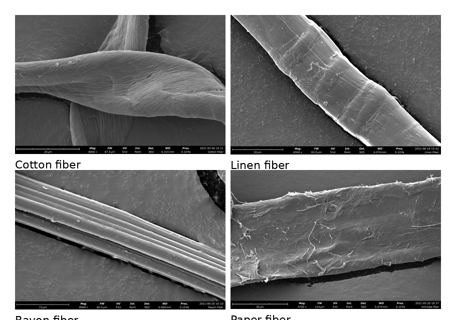
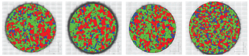
The cellulosic-fiber example illustrates the distinction between identification and root cause. IR spectroscopy can show that a contaminant is cellulosic, but SEM morphology can help distinguish cotton, linen, rayon, and paper-like sources. In formulated products, Raman imaging can add a second root-cause dimension by showing whether components are distributed consistently or whether compression, coating, or formulation differences created a spatially localized problem.
| Technique or platform | Information produced | Why it matters |
|---|---|---|
| Optical and digital microscopy | Visual morphology, dimensions, surface features, color, layering, and sample-selection context. | Documents the evidence before destructive testing and helps select specific particles or regions for analysis. |
| Raman microscopy and chemical mapping | Molecular fingerprints and spatial distribution of many APIs, excipients, pigments, polymers, and crystalline components. | Useful for suspect-versus-authentic comparisons, coating/core analysis, layered systems, and localized unknowns. |
| FTIR and IR microspectroscopy | Polymer, organic, excipient, adhesive, fiber, film, and residue identification. | Often strong for particles, fibers, packaging materials, cap liners, label adhesives, and contact-material comparisons. |
| SEM/EDX | High-resolution morphology plus elemental composition and elemental maps. | Critical for inorganic particles, fillers, talc-related signals, metals, corrosion, pigments, and source comparisons. |
| LC/MS, GC/MS, chromatography, NMR, or ICP-MS | Targeted or investigative molecular, volatile/semi-volatile, structural, or trace-element information. | Added when direct microanalysis is not enough or when confirmation, quantitation, or structural assignment is required. |
Establish the chemical and physical identity of a particle, residue, fiber, film, deposit, or mixed material before testing source hypotheses.
Determine the structure and formation pathway of an unknown peak, process impurity, degradation product, or drug-product reaction product.
Compare bulk or localized elemental evidence with raw materials, equipment, catalysts, packaging, and environmental sources.
Design sampling, blank, and sensitivity controls when the suspected cause is present only as a weak, sparse, or matrix-interfered signal.
Begin with careful documentation, sample preservation, and visual examination, then progressively narrow the contaminant's identity, source, and introduction mechanism. Use complementary techniques such as microscopy, spectroscopy, elemental analysis, thermal analysis, chromatography, and diffraction as appropriate. Conclusions should integrate multiple results to establish composition, physical form, probable origin, and potential product-quality impact.
Dissolution failures often arise from interacting changes in particle size, solid form, amorphous content, granulation, compression, lubrication, moisture exposure, excipient variability, coating, storage, or manufacturing conditions. Investigations should evaluate both formulation and process factors using orthogonal analytical methods rather than assuming the API is solely responsible.
Preserve the evidence and combine microscopy with spectroscopy, elemental analysis, diffraction, thermal analysis, and chromatography as appropriate. Foreign particulates may originate from equipment, raw materials, packaging, the environment, personnel, or product degradation. Analytical findings should be compared with manufacturing records and suspected-source materials to establish a defensible root cause and support effective corrective and preventive actions.
Potential causes include oxidation, photodegradation, hydrolysis, Maillard reactions, trace-metal catalysis, excipient incompatibility, solid-form changes, moisture uptake, coating degradation, microbial contamination, and temperature or humidity exposure. Because different mechanisms can produce similar color changes, use visual examination, spectroscopy, chromatography, thermal analysis, microscopy, and solid-state characterization to identify the mechanism and assess product-quality impact.
Determine what crystallized, when nucleation began, and which changes in composition, temperature, solvent environment, shear, moisture, or process timing created favorable conditions. Examine the crystals using microscopy, XRPD, Raman or FTIR spectroscopy, thermal analysis, particle characterization, and chromatography, then correlate the results with process data. The root cause may be several modest changes that collectively shifted the process into a crystallization-prone region.
Scale-up changes mixing, heat transfer, shear, drying, compression, residence time, material movement, and environmental exposure. Raw-material variability, longer hold times, equipment geometry, sampling limitations, and tighter timelines can expose weaknesses not apparent at laboratory or pilot scale. Failures often indicate that critical material attributes, the process design space, or scale-dependent relationships were not fully defined during development.
They determine what changed, where and when it changed, and why. Microscopy, spectroscopy, diffraction, thermal analysis, chromatography, particle characterization, and materials science are integrated with manufacturing records, process history, and formulation knowledge. The objective is to identify the physical or chemical mechanism behind the failure and develop corrective actions that improve process robustness.
Start by defining the scientific question, physical form of the evidence, and most likely type of change. Chromatography evaluates purity and degradation; XRPD identifies crystalline phases; Raman and FTIR establish molecular identity; microscopy reveals morphology and spatial distribution; thermal analysis evaluates transitions and volatile loss; particle methods characterize size and shape. Use a staged, minimally destructive strategy and complementary methods to confirm the failure mechanism.

Share the observed failure, affected and acceptable lots, process step and timeline, sample and storage history, prior analytical data, suspected sources, available comparison materials, and the hypotheses the investigation must test.