
Root Cause Investigations
Use the contaminant identity, morphology, location, and comparison results to test process, packaging, raw-material, or environmental source hypotheses.

Unknown-contaminant work starts with the physical evidence: what was found, where it was found, how it was collected, whether it was handled or isolated, and what comparison materials are available. A useful answer is rarely just a library hit. The goal is to identify the material class or composition well enough to support an investigation, then determine whether the analytical evidence is consistent with a proposed source.
Triclinic uses orthogonal workflows because visible particles, films, residues, fibers, and mixed debris often contain multiple components. Microscopy defines morphology and context; Raman and FTIR provide molecular identity; SEM/EDX adds elemental and spatial evidence; and chromatographic or MS methods can be added when the unknown is soluble, trace-level, degradant-like, or not suitable for direct microanalysis.
Fiber contaminants are common in pharmaceutical, cosmetic, food, packaging, and manufacturing environments. The practical question is not just whether a particle is cellulosic, but whether the evidence can distinguish cotton, linen, rayon, paper, filter media, wipe material, packaging debris, or another realistic source candidate.
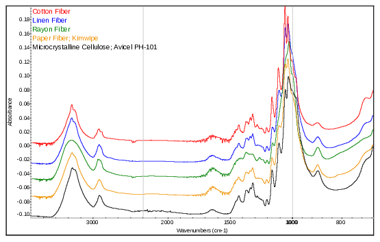
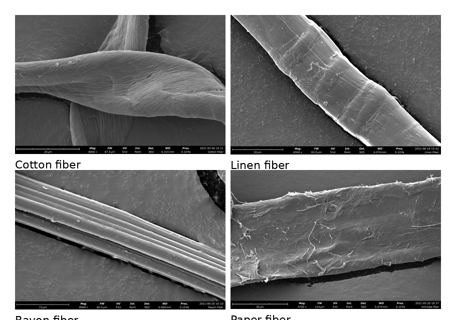
The cellulosic-fiber application note demonstrates why a single technique may not be enough. IR microspectroscopy classified cotton, linen, rayon, and paper fibers as cellulose-based materials, but the spectra were too similar for confident fiber-type discrimination. SEM then provided morphology - twisting, nodes, ridges, branching, and surface texture - that allowed the cellulosic fibers to be differentiated.
| Technique or platform | Information produced | Why it matters |
|---|---|---|
| Optical and digital microscopy | Visual morphology, dimensions, surface features, color, layering, and sample-selection context. | Documents the evidence before destructive testing and helps select specific particles or regions for analysis. |
| Raman microscopy and chemical mapping | Molecular fingerprints and spatial distribution of many APIs, excipients, pigments, polymers, and crystalline components. | Useful for suspect-versus-authentic comparisons, coating/core analysis, layered systems, and localized unknowns. |
| FTIR and IR microspectroscopy | Polymer, organic, excipient, adhesive, fiber, film, and residue identification. | Often strong for particles, fibers, packaging materials, cap liners, label adhesives, and contact-material comparisons. |
| SEM/EDX | High-resolution morphology plus elemental composition and elemental maps. | Critical for inorganic particles, fillers, talc-related signals, metals, corrosion, pigments, and source comparisons. |
| LC/MS, GC/MS, chromatography, NMR, or ICP-MS | Targeted or investigative molecular, volatile/semi-volatile, structural, or trace-element information. | Added when direct microanalysis is not enough or when confirmation, quantitation, or structural assignment is required. |
Use the contaminant identity, morphology, location, and comparison results to test process, packaging, raw-material, or environmental source hypotheses.
Measure bulk or localized elemental composition when an inorganic, metallic, mineral, catalyst, or corrosion-related source is suspected.
Plan sampling, isolation, blanks, and sensitivity controls when the unknown is sparse, microscopic, or difficult to recover.
Compare suspect products, dosage forms, packaging, seals, labels, and formulation signatures against authentic references.
Use optical microscopy, electron microscopy, and localized elemental mapping to characterize particles, fibers, residues, inclusions, and suspected contaminant sources.
Often. Microscopy-guided sampling can isolate selected particles or regions for Raman spectroscopy, Fourier-transform infrared spectroscopy, or scanning electron microscopy with energy-dispersive X-ray spectroscopy. Mixed materials may require multiple techniques and component-specific conclusions.
Yes. Retained lots, raw materials, packaging, process-contact materials, filters, cleaning agents, environmental samples, and supplier materials can be compared. The report should state whether the evidence supports, excludes, or remains inconclusive for each proposed source.
The report distinguishes confirmed identity, material-class assignment, evidence-consistent source comparison, and unresolved possibilities. It also states the analytical limitations and any additional work needed for source attribution or confirmation.
Provide photographs, collection method, location, product and process history, handling history, storage conditions, suspected sources, comparison materials, prior data, and the quality or manufacturing decision the analysis must support.

Share photographs, the collected unknown, good and suspect lots, collection and handling history, process location, suspected sources, available comparison materials, prior data, and the quality or manufacturing decision the identification must support.